Fasciola hepatica (the sheep liver fluke)
The common name of this parasite, the "sheep liver fluke," is somewhat misleading since this parasite is found in animals other than sheep (including cattle and humans), and the parasite resides in the bile ducts inside the liver rather than the liver itself. This species is a common parasite of sheep and cattle and, therefore, relatively easy to obtain. Thus, in introductory biology or zoology courses, it is often used as "THE" example of a digenetic trematode. This species has been studied extensively by parasitologists, and probably more is known about this species of digenetic trematode than any other.
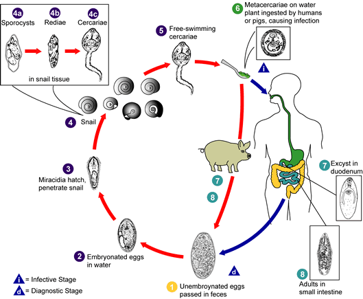
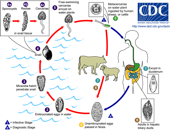
The adult parasites reside in the intrahepatic bile ducts, produce eggs, and the eggs are passed in the host's feces. After passing through the first intermediate host (a snail), cercariae encyst on vegetation. The definitive host is infected when it eats the contaminated vegetation. The metacercaria excysts in the definitive host's small intestine, and the immature worm penetrates the small intestine and migrates through the abdominal cavity to the host's liver. The juvenile worm penetrates and migrates through the host's liver and finally ends up in the bile ducts. The migration of the worms through the host's liver, and the presence of the worms in the bile ducts, are responsible for the pathology associated with fascioliasis.
Fasciola hepatica is found in parts of the United States, as well as in Great Britian, Ireland, Europe, the Middle East, the Far East, Africa, and Australia. Fascioliasis in sheep and cattle results in animals that show low productivity (low weight gain, low milk production, etc.). Also, in many countries, livers from animals infected with F. hepatica are condemned as unsuitable for human consumption. This not only results in a significant economic loss to ranchers and farmers, but it also results in the loss of an important source of protein. The infection can be diagnosed by finding eggs in the feces of animals, but the eggs are difficult to differentiate from closely related species such as Fasciolopsis buski. Several immunological methods for diagnosis are available.
| |
|
|
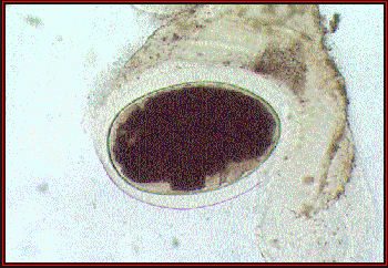
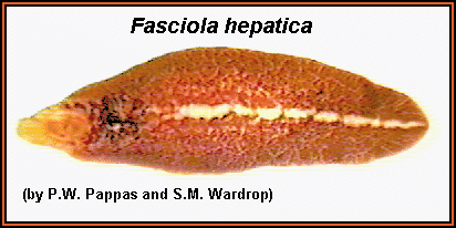
A stained whole mount of an adult Fasciola hepatica; approximate length = 20 mm. The internal organs of this species are characteristically highly branched, thus making it very difficult to differentiate the various internal organs.
|
| |
|
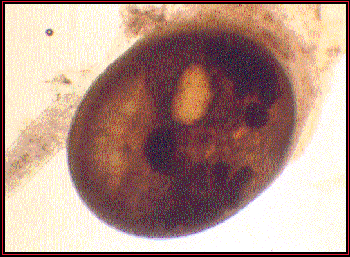
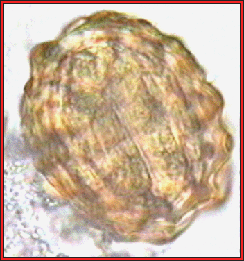
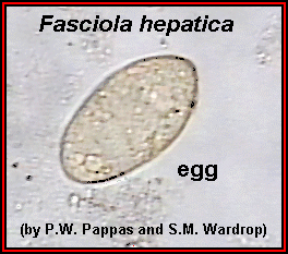 Fasciola hepatica egg; approximate size = 140 µm in length. Fasciola hepatica egg; approximate size = 140 µm in length.
|
Another example of a Fasciola hepatica egg, showing the operculum.
|
Life Cycle of the Fasciola hepatica
Fasciolopsis buskii
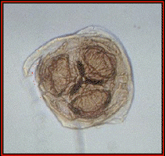
Fasciolopsis buski lives in the small intestine of humans and pigs. Measuring up to 80 mm in length, it is one of the largest trematodes found in humans. This parasite is found in many countries in the Orient and, as with many other parasites that infect humans, pigs serve as a reservoir host.
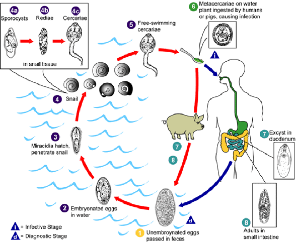
The life cycle of this parasite is similar to that of Fasciola hepatica. The worms produce eggs (up to 25,000 eggs per worm per day) that are passed in the host's feces. The first intermediate host is a snail, and the cercariae that emerge from the snail encyst on vegetation. Humans are infected with then eat vegetation contaminated with metacercariae.
Life cycle of the Fasciolopsis buski
Chronic infections with this parasite lead to inflammation, ulceration, hemorrhage, and abscesses of the small intestine, and these can ultimately lead to the host's death. Diagnosis of the disease is based on recovering eggs in the host's feces.
Several books and a number of web sites state that this parasite either causes directly or is associated with an increased risk of cancer, HIV, or any number of other diseases in humans. There is absolutely no evidence whatsoever that this parasite causes cancer, HIV, or any other disease in humans.
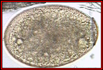
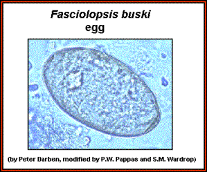 Fasciolopsis buski egg. The egg is very similar to that of Fasciola hepatica; approximate size = 130 µm in length.
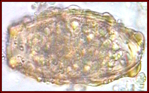
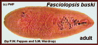 Stained whole mount of a Fasciolopsis buski adult; approximate length = 50 mm.
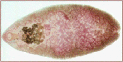
Another example of an adult Fasciolopsis buski. This specimen is stained lighter and is much larger than the example above, and some organs are seen more easily. (Original image from Taipei Medical College Parasitology web site, and modified for use.
These wonderful images are provided to you by: www.biosci.ohio-state.edu
They an extensive library of information on over 300 types of parasites!
Ascaris lumbricoides and Ascaris suum
(intestinal roundworms of humans and pigs)
 Ascaris lumbricoides is one of the largest and most common parasites found in humans. The adult females of this species can measure up to 18 inches long (males are generally shorter), and it is estimated that 25% of the world's population is infected with this nematode. The adult worms live in the small intestine and eggs are passed in the feces. A single female can produce up to 200,000 eggs each day! About two weeks after passage in the feces the eggs contain an infective larval or juvenile stage, and humans are infected when they ingest such infective eggs. The eggs hatch in the small intestine, the juvenile penetrates the small intestine and enters the circulatory system, and eventually the juvenile worm enters the lungs. In the lungs the juvenile worm leaves the circulatory system and enters the air passages of the lungs. The juvenile worm then migrates up the air passages into the pharynx where it is swallowed, and once in the small intestine the juvenile grows into an adult worm. Why Ascaris undergoes such a migration through the body to only end up where it started is unknown. Such a migration is not unique to Ascaris, as its close relatives undergo a similar migration in the bodies of their hosts (view diagram of the life cycle below). Ascaris lumbricoides is one of the largest and most common parasites found in humans. The adult females of this species can measure up to 18 inches long (males are generally shorter), and it is estimated that 25% of the world's population is infected with this nematode. The adult worms live in the small intestine and eggs are passed in the feces. A single female can produce up to 200,000 eggs each day! About two weeks after passage in the feces the eggs contain an infective larval or juvenile stage, and humans are infected when they ingest such infective eggs. The eggs hatch in the small intestine, the juvenile penetrates the small intestine and enters the circulatory system, and eventually the juvenile worm enters the lungs. In the lungs the juvenile worm leaves the circulatory system and enters the air passages of the lungs. The juvenile worm then migrates up the air passages into the pharynx where it is swallowed, and once in the small intestine the juvenile grows into an adult worm. Why Ascaris undergoes such a migration through the body to only end up where it started is unknown. Such a migration is not unique to Ascaris, as its close relatives undergo a similar migration in the bodies of their hosts (view diagram of the life cycle below).
Ascaris infections in humans can cause significant pathology. The migration of the larvae through the lungs causes the blood vessels of the lungs to hemorrhage, and there is an inflammatory response accompanied by edema. The resulting accumulation of fluids in the lungs results in "ascaris pneumonia," and this can be fatal. The large size of the adult worms also presents problems, especially if the worms physically block the gastrointestinal tract. Ascaris is notorious for its reputation to migrate within the small intestine, and when a large worm begins to migrate there is not much that can stop it. Instances have been reported in which Ascaris have migrated into and blocked the bile or pancreatic duct or in which the worms have penetrated the small intestine resulting in acute (and fatal) peritonitis. Ascaris seems to be especially sensitive to anesthetics, and numerous cases have been documented where patients in surgical recovery rooms have had worms migrate from the small intestine, through the stomach, and out the patient's nose or mouth.
Ascaris suum is found in pigs. Its life cycle is identical to that of A. lumbricoides. If a human ingests eggs of A. suum the larvae will migrate to the lungs and die. This can cause a particularly serious form of "ascaris pneumonia." Adult worms of this species do not develop in the human's intestine. (Some parasitologists believe that there is but one species of Ascaris that infects both pigs and humans, but any commentary on this issue is beyond the scope of this web site.)
Infections of Ascaris are diagnosed by finding characteristic eggs in the feces of the infected host.
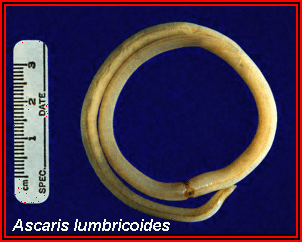
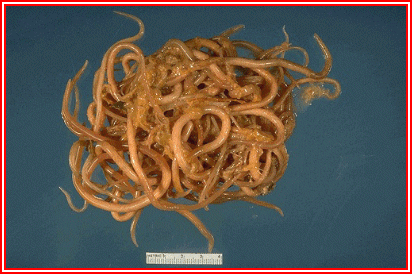 A large mass of Ascaris lumbricoides that was passed from the intestinal tract. The ruler at the bottom of the image is 4 cm (about 1.5 inches) in length.
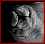
An en face view of Ascaris. Note the presence of three large lips, a characteristic of all ascarids. (Original image from Oklahoma State University, College of Veterinary Medicine.)
A scanning electron micrograph of the anterior end of Ascaris showing the three prominent "lips." (Original image from "Wormland".)
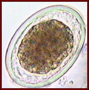
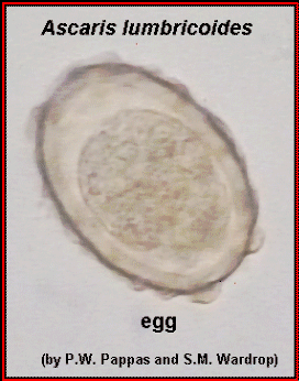 Ascaris lumbricoides, fertilized egg. Note that the egg is covered with a thick shell that appears lumpy (bumpy) or mammillated; approximate size = 65 µm in length.
 Another example of a fertilized Ascaris lumbricoides egg. (Original image from: Atlas of Medical Parasitology.)
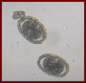
 An example of an unfertilized A. lumbricoides egg. (Original image from: Atlas of Medical Parasitology.)
 A "decorticated," fertilized, Ascaris lumbricoides. (Original image from: Atlas of Medical Parasitology.)
 Eggs of Ascaris suum. A. suum is a common parasite of pigs. The eggs are virtually indistinguishable from those of A. lumbricoides. (Original image from Oklahoma State University, College of Veterinary Medicine.)
 A female Ascaris lumbricoides. Females of this species can measure over 16 inches long. This specimen was passed by a young girl in Florida. (Original image from DPDx [Identification and Diagnosis of Parasites of Public Health Concern].)
 Female and male Ascaris lumbricoides; the female measures approximately 16 inches (40 cm) in length.
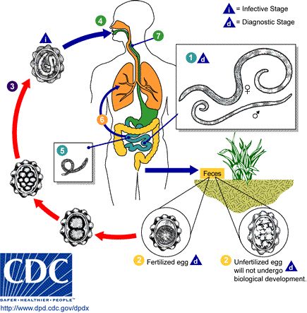
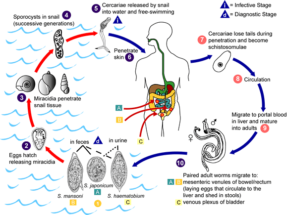
Adult Ascaris lumbricoides
worms (1) live in the lumen of the small
intestine. A female may produce 200,000 eggs each day, which
are passed with the feces (2) of the host.
Ingested unfertilized eggs are not infective, but fertile
eggs begin to develop and become infective after 18 days to
several weeks (3), depending on
environmental conditions (an optimal environment being
moist, warm, shaded soil). After infective eggs are
swallowed (4), the larvae hatch (5),
invade the intestinal mucosa, and are carried via first the
portal and then the systemic circulation to the lungs
(6). The larvae mature further in the lungs
for 10 to 14 days, then penetrate the alveolar walls, ascend
the bronchial tree to the throat, and are swallowed
(7). Upon reaching the small intestine, they
develop into adult worms (1). Between two
and three months are required from ingestion of infective
eggs to oviposition (egg-laying) by the adult female. Adult
worms can live one to two years.
These images are provided to you by:www.biosci.ohio-state.edu
Back to Parasite Cleanse
Page
.png)
Parasite
Cleanse by Dr. Clark
|