"Electron Therapy Research"
 -
by John Crane -
by John Crane
One of Royal Rife's
research partners, and developer of the first modern day
frequency generator for home use... The RPG1000.
And later, the RPG900. (Rife Programmable
Generator) These were the first "contact pad"
instruments, which most of today's frequency generators are
fashioned after.
"This report on Electron Therapy is primarily concerned with the detection and cure of cancer and related diseases. It is presented for research only.
It is a matter of record that, while orthodox medical and surgical methods have had limited success with cancer discovered in the early stage, they have had little or no success with treatment of the more advanced cases. The cause of cancer is still listed in some of the current medical textbooks as unknown.
Since research to determine the cause and effect a cure of cancer is being carried out in many and various directions, it seems reasonable to explore every possible avenue toward a true understanding of the cause of cancer and hence, its proper treatment This report concerns itself with the discovery of a virus causing cancer and a method of treatment which excited considerable interest among reputable doctors and laboratory specialists as long ago as 1934. This theory has stood up under hundreds of controlled laboratory tests and has been applied to treat human cancer in dozens of cases.
Rife Virus Microscope Institute, in conjunction with various doctors throughout the United States and Canada, is at the present time (1960-ed.) engaged in carrying out a controlled series of tests in order to re-establish the finding of Dr. Royal R. Rife in his life study of the cause and cure of cancer.
Dr. Royal Raymond Rife first conceived the idea of the Frequency Instrument in conjunction with his work in developing the Rife Universal Microscope. Since the determining of the cancer requires the high power of the Rife Microscope, a brief description of the construction and theory of operation of the Rife Microscope will be included in this report.
THE UNIVERSAL MICROSCOPE
Dr. Royal R. Rife over a period of 30 years designed and built in his own laboratory five microscopes of power and resolution far beyond the so-called law of optical physics. In their power magnification these instruments vary from 9,000 to
60,000 times, far beyond the limits of the standard research instrument. The commercial microscope being manufactured today is inadequate for the observation of filterable viruses of disease (as these minute, live, living entities are less that 1/20 of one micron in dimension). Thus the need for a device which would carry us farther into this important field of endeavor. We will describe in some detail the most powerful of these microscopes, known as the Universal Microscope.
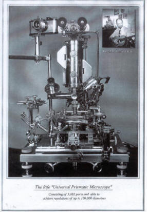
The Universal Microscope, which is the largest and most powerful of the light microscopes, developed in 1933, consists of 5,682 parts.
This microscope derives its name from its adaptability to all fields of microscopical work. The microscope is fully equipped with separate substage condenser units for transmitted and mono chromatic beam, darkfield, polarized, and slit-ultra illumination, and includes a special device for crystallography. The entire optical system of lenses and prisms as well as the illuminating units are made of block-crystal quartz.
The illuminating unit used for the examination of the filterable form of disease organisms contains 14 lenses and prisms, three of which are in the hi-intensity incandescent lamp, four in the Risley prism, and seven in the achromatic condenser system. Two circular, wedge-shaped prisms are suspended between the source of light and the specimen being examined. The two prisms are used for changing the angle of incidence of the light passing through the specimen being examined. When the light passes through these prisms, it is divided or split into two beams, one of which is refracted to such an extent that it is reflected to the side of the prism while the second beam is permitted to pass through the prism and illuminate the specimen owing to its chemical constituents.
The mounting arrangement on the Universal Microscope permits each of the two prisms to be rotated in opposite directions by a vernier control throughout 360 degrees. This vernier adjustment permits bending the transmitted beam of light at variable angles of incidence while, at the same time, a small portion of the spectrum is projected into the axis of the microscope owing to the chemical constituents of the microorganism. The vernier adjustment permits only a small portion of the spectrum to be visible at any one time, but it is possible to select any portion from one end of the spectrum to the other. When that portion of the spectrum is reached where both the organism and the color band vibrate in exact accord, a definite characteristic spectrum is emitted by the organism.
PRINCIPLE OF PARALLEL RAYS
In the case of the filter-passing form of the Bacillus Typhosus, a turquoise blue color is emitted and the plane of polarization deviated plus 4.8 degrees. The predominating chemical constituents of the organism are next ascertained after which the quartz prisms are adjusted by means of the vernier control to minus 4.8 degrees so the opposite angle of refraction may be obtained. A monochromatic beam of light corresponding exactly to the frequency of the organism is then passed through the specimen along with the direct transmitted light. This beam permits the observer to view the organism stained in its true chemical color and reveals its own individual structure in a field which is brilliant with light.
The rays of light refracted by the specimen enter the objective lens and are carried up the tube in parallel rays through twenty-one light bends to the ocular lens. A tolerance of less that one wave length of visible light is permitted in the core beam of illumination. Iii the standard optical microscope, the light rays tend to converge as they rise higher and finally cross each other, arriving at the ocular lens, separated by a considerable distance.
This microscope derives its name from its adaptability to all fields of microscopical work. The microscope is fully equipped with separate substage condenser units for transmitted and mono chromatic beam, darkfield, polarized, and slit-ultra illumination, and includes a special device for crystallography. The entire optical system of lenses and prisms as well as the illuminating units are made of block-crystal quartz.
The illuminating unit used for the examination of the filterable form of disease organisms contains 14 lenses and prisms, three of which are in the hi-intensity incandescent lamp, four in the Risley prism, and seven in the achromatic condenser system. Two circular, wedge-shaped prisms are suspended between the source of light and the specimen being examined. The two prisms are used for changing the angle of incidence of the light passing through the specimen being examined. When the light passes through these prisms, it is divided or split into two beams, one of which is refracted to such an extent that it is reflected to the side of the prism while the second beam is permitted to pass through the prism and illuminate the specimen owing to its chemical constituents.
The mounting arrangement on the Universal Microscope permits each of the two prisms to be rotated in opposite directions by a vernier control throughout 360 degrees. This vernier adjustment permits bending the transmitted beam of light at variable angles of incidence while, at the same time, a small portion of the spectrum is projected into the axis of the microscope owing to the chemical constituents of the microorganism. The vernier adjustment permits only a small portion of the spectrum to be visible at any one time, but it is possible to select any portion from one end of the spectrum to the other. When that portion of the spectrum is reached where both the organism and the color band vibrate in exact accord, a definite characteristic spectrum is emitted by the organism.
The Rife microscopes, as the rays are about to cross each other, a specially designed quartz prism is inserted which serves to separate the light rays to a near parallel line again. Additional prisms are inserted each time the rays are ready to cross.
These prisms, located in the tube, are adjusted and held in alignment by micrometer? screws in special tracks made of magnelium a metal having the closest expansion coefficient of any metal to quartz. These prisms are separated by a distance of only 30 millimeters. Thus, the greatest distance that the image in the Universal Microscope is projected through any one media, either quartz or air, is 30 millimeters instead of the 160 to 190 millimeters employed in the air-filled type of the ordinary microscope.
It is this principle of parallel rays used in the Universal Microscope and the resultant shortening of projection distance between any two blocks or prisms plus the fact that objective lenses can thus be substitutes for oculars, (these oculars being three matched pairs of lO-mm, 7-mm and 4-mm objectives in short mounts) which make possible not only the unusually high magnification and resolution but which serve to eliminate virtually all chromatic and spherical aberration.
The universal stage is a double rotating stage graduated through 360 degrees in quarter-minute arc divisions. The upper segment carries the mechanical stage having a movement of 40 degrees, and the body assembly which can move horizontally over the condenser and provide an angular tilt of 40 degrees + or -.
The microscope stands 24 inches high and weighs 200 pounds. The base is composed of cast nickel-steel plate, accurately surfaced and equipped with three leveling screws and two spirit levels set at 90 degrees. The course adjustment, a clock thread screw with 40 threads to the inch, slides in a 11/4 dovetail which gibs directly onto the pillar post. The stage, in conjunction with a hydraulic lift, acts as a lever in operating the fine adjustment.
A 6-guage screw, having 100 threads to the inch, is worked through a gland into a hollow glycerine-filled post, the glycerine being displaced and replaced at will as the screw is turned clockwise or counterclockwise allowing a 5 to 1 ratio on the lead screw. This hydraulic action assures complete absence of drag or inertia.
The fine adjustment being 700 times wore sensitive than the ordinary microscopes, requires a length of time from ten minutes to one-half hour to focus. This time at first glance seems a disadvantage, but it is felt that, for the overall results obtained, the time required is only a slight inconvenience compared to the many years' research and the actual results obtained in isolating and looking upon disease-causing organisms in their true form (to be continued)
A 45 minute video (VHS) of Royal Raymond Rife in his laboratory is available from BSRF and this tape shows the inflicting of cancer in laboratory mice, their cure with Rife's ray tube frequency instruments. It is transferred from a 16mm film and is narrated by John Crane. This film is proof that the cure for cancer (one of many) has been covered up!
John Crane can be contacted at 4246 Pepper Drive; San Diego, CA 92105. He is dedicated to preserving and promoting the work of Royal Raymond Rife and the Rife Virus microscope Institute."
-End

Electron Therapy by John Crane
|